Nara Higano, PhD ’17, is using her knowledge of physics to help the smallest patients.
While some PhD-level physicists end up working at supercolliders, nuclear reactors, or university labs, Nara Higano, PhD ’17, has found her calling in what might seem like an unexpected place: the neonatal intensive care unit (NICU) at Cincinnati Children’s Hospital Medical Center.
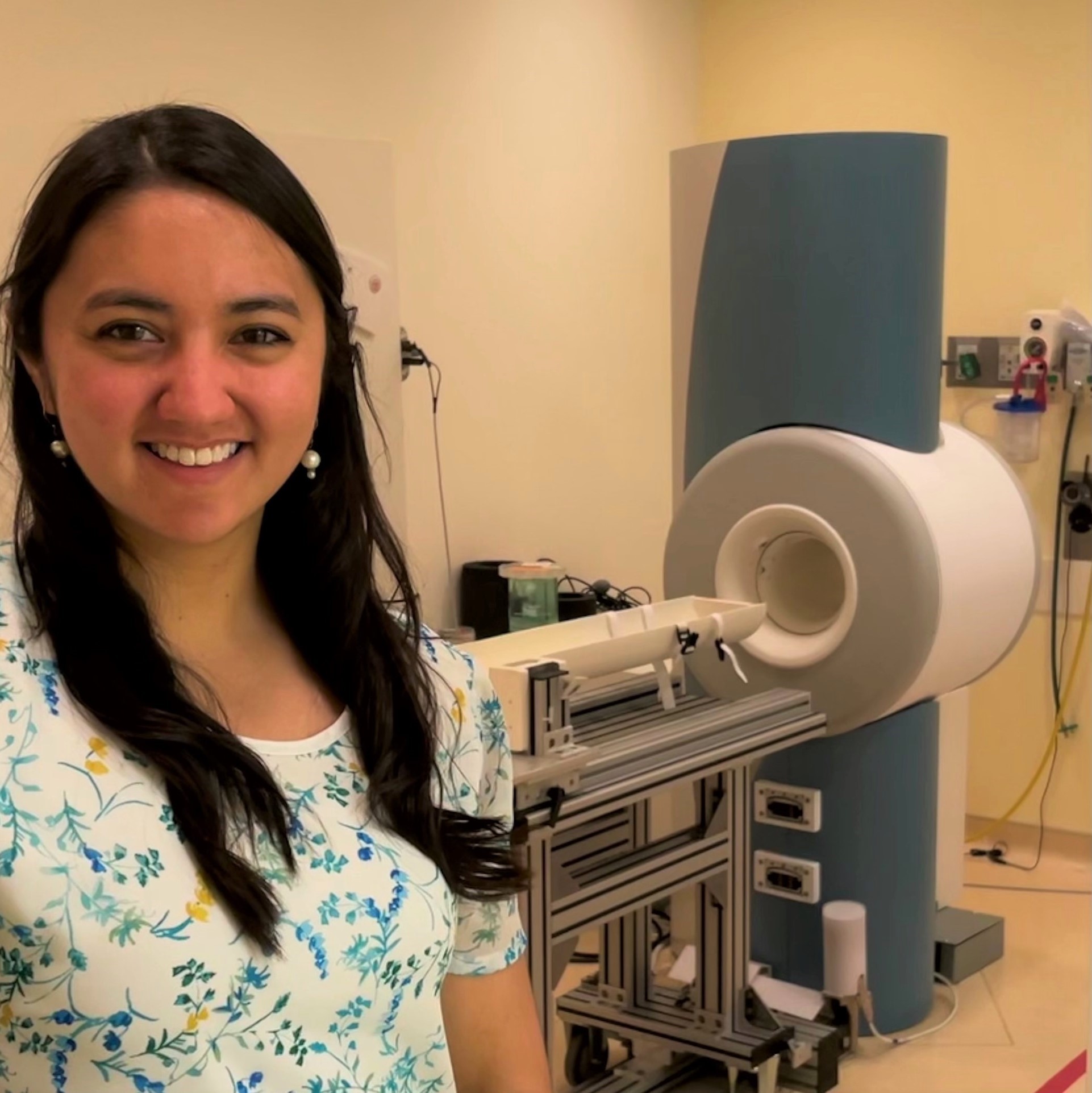
For the last five years, Higano has been using her knowledge of protons and electromagnetism to improve the lives of premature infants. Her work focuses on fine-tuning the use of magnetic resonance imaging (MRI) technology to view fragile lungs that haven’t fully developed. “MRIs can produce beautiful images, but we’ve come nowhere close to fully tapping their potential in medical settings, including the NICU,” she said. “I’m using the physics I learned in my PhD studies at WashU to help sick babies.”
MRI machines use a combination of magnetic fields and radio waves to create images. But what’s more important than what they use is what they leave out: potentially harmful ionizing radiation. Other options, like computed tomography (CT) scans, can produce exquisitely detailed images, but they are rarely used in NICUs, in part because the dose of ionizing radiation produces risks that generally outweigh any potential benefits. And while X-rays produce a relatively small amount of radiation, the images aren’t crisp enough to display the subtle nuances of diseased lung tissues. All of these reasons make MRIs an appealing option for fragile patients who may need to be scanned many times throughout their lives.
Getting an image with an MRI isn’t as simple as pushing a button on a camera. The image depends on precise control of spinning protons within magnetic fields, which are intentionally changed over time and space. And that’s where Higano and other physicists fit into the picture. “The image of a person’s body is only possible because someone manipulated nuclear spins,” Higano said. “In order for MRIs to be used in more settings, someone will have to think about the physics involved.”
Higano got her bachelor’s degree in physics at Gustavus Adolphus College in Saint Peter, Minnesota, but she didn’t see the full possibilities of the field until she got to WashU for graduate school. Alongside her courses in theoretical physics, relativity, and quantum mechanics, she learned about the use of physics in medicine. “Some of the researchers in the physics department were working closely with investigators at the med school, and I got really interested in medical physics,” she said.
As a grad student, she worked as a teaching assistant for the popular Physics of the Heart undergraduate course taught by James G. Miller, now the Albert Gordon Hill Professor Emeritus of Physics. “I was essentially taking the course along with the undergrads, and I really became enthralled with how physics can be applied to biological systems,” she said. “I started to really appreciate the potential for less traditional applications of my physics education.”
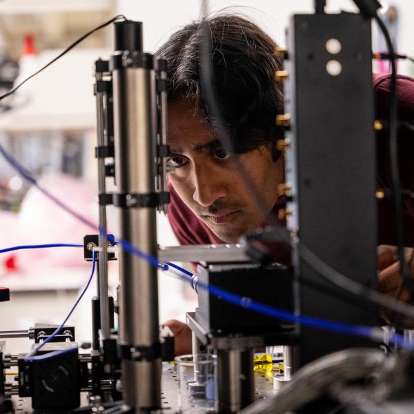
Higano deepened her understanding of medical physics working in the lab of her thesis co-advisor, Mark Conradi, now a professor emeritus of physics. Conradi’s work involved using xenon gas to view lung disease in a novel way. In order to do its job, the gas has to be “hyperpolarized” using a combination of high-powered lasers and magnets — a type of manipulation of quantum mechanical states that is being championed by the Center for Quantum Leaps, a signature initiative of the Arts & Sciences Strategic Plan. “You have to marry several principles of physics to make it work,” Higano said.
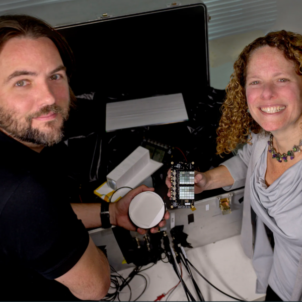
At Cincinnati Children’s Hospital, Higano is part of a team dedicated to unlocking the potential of MRIs in the NICU. Physicists, engineers, and physical chemists work together to generate the types of images that can identify clinical problems and guide treatments. “We are super interdisciplinary and we work on a nearly daily basis with the physicians who keep our work grounded in key medical questions that will actually help patients,” Higano said.
The team is motivated by the hope that sharper images could add clarity to clinical decisions. Physicians, for instance, could potentially use lung MRIs to identify the premature infants who are most likely to respond to corticosteroids.
Despite their potential, MRIs are still a largely untapped resource for treating premature infants. “There are only a few centers in the world that have the logistics to bring MRIs to the NICU,” Higano said. “It takes a huge team effort to move forward with this sort of imaging.”
That makes Higano’s work all the more valuable. In 2020, she was a lead author of a first-of-its-kind study that used hyperpolarized gas MRIs to compare the space in the air sacs of healthy infants and those with lung disease. She hopes that demonstrating the power and accuracy of the procedure can help it reach more NICUs. “We would love to be able to capitalize on the work our center has done to really demonstrate the clinical relevance of these techniques,” she said.
The bustle and high stakes of a NICU may seem far removed from a physics classroom, but Higano says her training in quantum physics and electromagnetism at WashU has served her well in her current role. “I’m able to provide a crucial piece to the puzzle,” she said. “It’s amazing to be able to connect the dots in a way that’s going to actually impact somebody’s lifelong health.”